What is a head MRI?
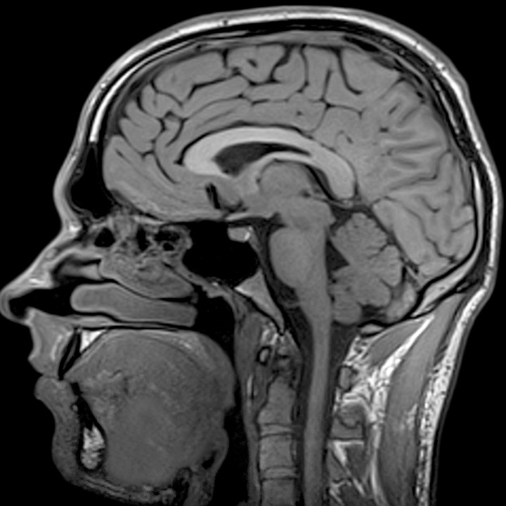
Magnetic resonance imaging (MRI) of the head is a painless, noninvasive test that produces detailed images of your brain and brain stem. An MRI machine creates the images using a magnetic field and radio waves. This test is also known as a brain MRI or a cranial MRI. You will go to a hospital or radiology center to take a head MRI.
An MRI scan is different from a CT scan or an X-ray in that it doesn’t use radiation to produce images. An MRI scan combines images to create a 3-D picture of your internal structures, so it’s more effective than other scans at detecting abnormalities in small structures of the brain such as the pituitary gland and brain stem. Sometimes a contrast agent, or dye, can be given through an intravenous (IV) line to better visualize certain structures or abnormalities.
Why do I need a head MRI?
A head MRI is a useful tool for detecting a number of brain conditions, including:
- aneurysms, or bulging in the blood vessels of the brain
- multiple sclerosis
- spinal cord injuries
- hydrocephalus, a buildup of spinal fluid in the brain cavities
- stroke
- infections
- tumors
- cysts
- swelling
- hormonal disorders, such as acromegaly and Cushing’s syndrome
- hemorrhage, or bleeding
- inflammation
- problems with development or structure (such as a Chiari malformation)
- blood vessel issues
- an issue due to a previous head injury
A head MRI can help determine whether you sustained any damage from a stroke or head injury. Your doctor may also order a head MRI to investigate symptoms such as:
- dizziness
- weakness
- seizures
- changes in thinking or behavior
- blurry vision
- chronic headaches
These symptoms may be due to a brain issue, which an MRI scan can help detect.
source: Healthline