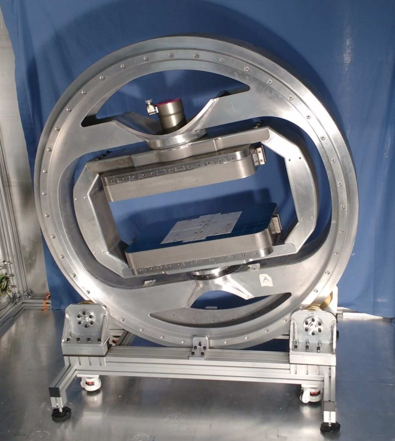
A new “mini-MRI” prototype has been developed by scientists at Imperial College London. The scanner is designed to improve how doctors diagnose and treat knee injuries. Research published on the new design mentions how the mini-MRI produces more accurate and crisper images of the anatomical structure of the knee versus a traditional MRI scan.
The scanner can potentially be used to diagnose sports injuries faster and more accurately which will allow athletes to start performing again sooner.
To develop the scanner, scientists decreased the size of their MRI machine by utilizing the “magic angle.” Researcher Dr. Karyn Chappell explains: “the brightness of these tissues such as tendons and ligaments in MRI images strongly depends on the angle between the collagen fibers and the magnetic field of the scanner. If this angle is 55 degrees, the image can be very bright, but for other angles it is usually very dark.”
The mini-MRI changes the orientation of the magnetic field much faster than traditional MRI machines. This produces higher quality images with lower field sizes.
Knee injuries in particular can be difficult to see and diagnose on a traditional MRI. Knee injuries commonly affect either the tendons, ligaments, or the meniscus which are not typically visible with MRI.
The mini-MRI changes all of that and will revolutionize how some of the most common injuries are diagnosed and treated.
Human trials are set for 2020.